The 3-Minute Rule for Circular Dichroism
Table of ContentsAn Unbiased View of Uv/visIndicators on Uv/vis You Should KnowLittle Known Facts About Circularly Polarized Luminescence.How Circular Dichroism can Save You Time, Stress, and Money.The Best Strategy To Use For Uv/visGetting My Uv/vis To WorkRumored Buzz on SpectrophotometersThe Basic Principles Of Uv/vis/nir Indicators on Spectrophotometers You Need To KnowThe 6-Second Trick For SpectrophotometersRumored Buzz on Uv/visNot known Incorrect Statements About Uv/vis Our Circular Dichroism PDFs
It is then scanned through the sample and the recommendation solutions. Portions of the event wavelengths are sent through, or shown from, the sample and the recommendation. The resultant light strikes the photodetector gadget, which compares the relative strength of the two beams. Electronic circuits transform the relative currents into direct transmission portions and/or absorbance/concentration values.The transmission of a referral substance is set as a baseline (information) value, so the transmission of all other compounds are recorded relative to the initial "zeroed" compound. The spectrophotometer then transforms the transmission ratio into 'absorbency', the concentration of specific parts of the test sample relative to the initial compound.
Because samples in these applications are not readily available in big quantities, they are particularly matched to being evaluated in this non-destructive technique. In addition, precious sample can be conserved by utilizing a micro-volume platform where just 1u, L of sample is required for complete analyses. A quick explanation of the treatment of spectrophotometry consists of comparing the absorbency of a blank sample that does not contain a colored substance to a sample which contains a colored substance.
3 Easy Facts About Uv/vis/nir Shown
In biochemical experiments, a chemical and/or physical residential or commercial property is chosen and the treatment that is used specifies to that residential or commercial property in order to derive more details about the sample, such as the quantity, purity, enzyme activity, and so on. Spectrophotometry can be utilized for a number of strategies such as identifying optimum wavelength absorbance of samples, determining ideal p, H for absorbance of samples, identifying concentrations of unidentified samples, and identifying the p, Ka of various samples.: 21119 Spectrophotometry is likewise a useful procedure for protein filtration and can also be used as an approach to create optical assays of a compound.
It is possible to understand the concentrations of a 2 component mix utilizing the absorption spectra of the standard options of each component. To do this, it is needed to know the extinction coefficient of this mixture at 2 wave lengths and the extinction coefficients of solutions which contain the recognized weights of the two parts.
Some Known Details About Uv/vis/nir
Area. The concentration of a protein can be approximated by measuring the OD at 280 nm due to the presence of tryptophan, tyrosine and phenylalanine.
Nucleic acid contamination can also interfere. This approach needs a spectrophotometer capable of determining in the UV region with quartz cuvettes.: 135 Ultraviolet-visible (UV-vis) spectroscopy involves energy levels that delight electronic shifts. Absorption of UV-vis light thrills molecules that remain in ground-states to their excited-states. Visible region 400700 nm spectrophotometry is utilized extensively in colorimetry science.
20. 8 O.D. Ink makers, printing companies, textiles suppliers, and a lot more, need the data provided through colorimetry. They take readings in the area of every 520 nanometers along the noticeable area, and produce a spectral reflectance curve or a data stream for alternative presentations. These curves can be utilized to evaluate a brand-new batch of colorant to examine if it makes a match to specs, e.
The smart Trick of Uv/vis That Nobody is Talking About
Traditional noticeable area spectrophotometers can not find if a colorant or the base material has fluorescence. This can make it hard to manage color issues if for instance one or more of the printing inks is fluorescent. Where a colorant contains fluorescence, a bi-spectral fluorescent spectrophotometer is utilized (https://www.quora.com/profile/Julie-Ann-DeSa-Lorenz). There are two significant setups for visual spectrum spectrophotometers, d/8 (round) and 0/45.
Researchers use this instrument to measure the amount of substances in a sample. If the substance is more focused more light will be soaked up by the sample; within little varieties, the Beer, Lambert law holds and the absorbance in between samples vary with concentration linearly. In the case of printing measurements two alternative settings are frequently used- without/with uv filter to control much better the impact of uv brighteners within the paper stock.
The 2-Minute Rule for Circular Dichroism
Some applications require small volume measurements which can be performed with micro-volume platforms. As described in the applications section, spectrophotometry can be used in both qualitative and quantitative analysis of DNA, RNA, and proteins. Qualitative analysis can be used and spectrophotometers are utilized to tape spectra of substances by scanning broad wavelength regions to identify the absorbance homes (the strength of the color) of the substance at each wavelength.
The Only Guide to Circularly Polarized Luminescence
One significant aspect is the kind of photosensors that are available for different spectral regions, but infrared measurement is also difficult since practically whatever discharges IR as thermal radiation, specifically at wavelengths beyond about 5 m. Another complication is that quite a couple of materials such as glass and plastic absorb infrared, making it incompatible as an optical medium.
Samples for IR spectrophotometry may be smeared between 2 discs of potassium bromide or ground with potassium bromide and pushed into a pellet. Where liquid services are to be determined, insoluble silver chloride is utilized to build the cell. Spectroradiometers, which operate almost like the visible region spectrophotometers, are created to measure the spectral density of illuminants. 2013. p. 13. Allen, DW; Cooksey, C; Tsai, BK (Nov 13, 2009). "Spectrophotometry". Retrieved Dec 23, 2018. Ninfa AJ, Ballou DP, Benore M (2010 ). Fundamental Laboratory Techniques for Biochemistry and Biotechnology (second ed.). Hoboken: Wiley & Sons. ISBN 9780470087664. OCLC 488246403. Schwedt G (1997 ). The necessary guide to analytical chemistry.
Oke, J. B.; Gunn, J. E.
Excitement About Uv/vis
Ninfa AJ, Ballou DP, Benore M (2015 ). Fundamental Laboratory Techniques for Biochemistry and Biotechnology (3, rev. ed.). UV/Vis/NIR. Lab Equipment.
Some Known Details About Uv/vis
Retrieved Jul 4, 2018. Trumbo, Toni A.; Schultz, Emeric; Borland, Michael G.; Pugh, Michael Eugene (April 27, 2013). "Applied Spectrophotometry: Analysis of a Biochemical Mixture". Biochemistry and Molecular Biology Education. 41 (4 ): 24250. doi:10. 1002/bmb. 20694. PMID 23625877. (PDF). www. mt.com. Mettler-Toledo AG, Analytical. 2016. Obtained Dec 23, 2018. Cortez, C.; Szepaniuk, A.; Gomes da Silva, L.
"Checking Out Proteins Purification Methods Animations as Tools for the Biochemistry Mentor". Journal of Biochemistry Education. 8 (2 ): 12. doi:. Garrett RH, Grisham CM (2013 ). Biochemistry. Belmont, CA: Cengage. p. 106. ISBN 978-1133106296. OCLC 801650341. Vacation, Ensor Roslyn (May 27, 1936). "Spectrophotometry of proteins". Biochemical Journal. 30 (10 ): 17951803. doi:10. 1042/bj0301795.
PMID 16746224. Hermannsson, Ptur G.; Vannahme, Christoph; Smith, Cameron L. C.; Srensen, Kristian T.; Kristensen, Anders (2015 ). "Refractive index dispersion noticing utilizing a selection of photonic crystal resonant reflectors". Applied Physics Letters. 107 (6 ): 061101. Bibcode:2015 Ap, Ph, L. 107f1101H. doi:10. 1063/1. 4928548. S2CID 62897708. Mavrodineanu R, Schultz JI, Menis O, eds.
Uv/vis for Dummies
U.S. Department of Commerce National Bureau of Standards unique publication; 378. Washington, D.C.: U.S. National Bureau of Standards. p. 2. OCLC 920079.
The procedure begins with a controlled light that illuminates the analyzed sample. When it comes to reflection, as this light engages with the sample, some is absorbed or produced. The produced light journeys to the detector, which is analyzed, quantified, and presented as industry-standard color scales and indices.
All terms are examined over the visible spectrum from 400 to 700 nm. In the case of transmission, when the light communicates with the sample, it is either absorbed, shown, or transmitted.
The 5-Minute Rule for Circularly Polarized Luminescence
Examples include APHA (American Public Health Association) for watercolor and purity analysis, ASTM D1500 for petrochemical color analysis, edible oil indices utilized in food, and color analyses of beverages. The simplified mathematics looks like this:. Where T is the transmission coefficient. All terms are assessed over the visible spectrum from 400 to 700 nm.
Image Credit: Matej Kastelic/ Dr. Arnold visite site J. Beckman and his coworkers at the National Technologies Laboratories first invented the spectrophotometer in 1940. In 1935 Beckman founded the business, and the discovery of the spectrophotometer was their most ground-breaking creation.
Facts About Circular Dichroism Uncovered
Over time, researchers kept enhancing the spectrophotometer design to boost its performance. The UV capabilities of the design B spectrophotometer were improved by changing the glass prism with a quartz prism.
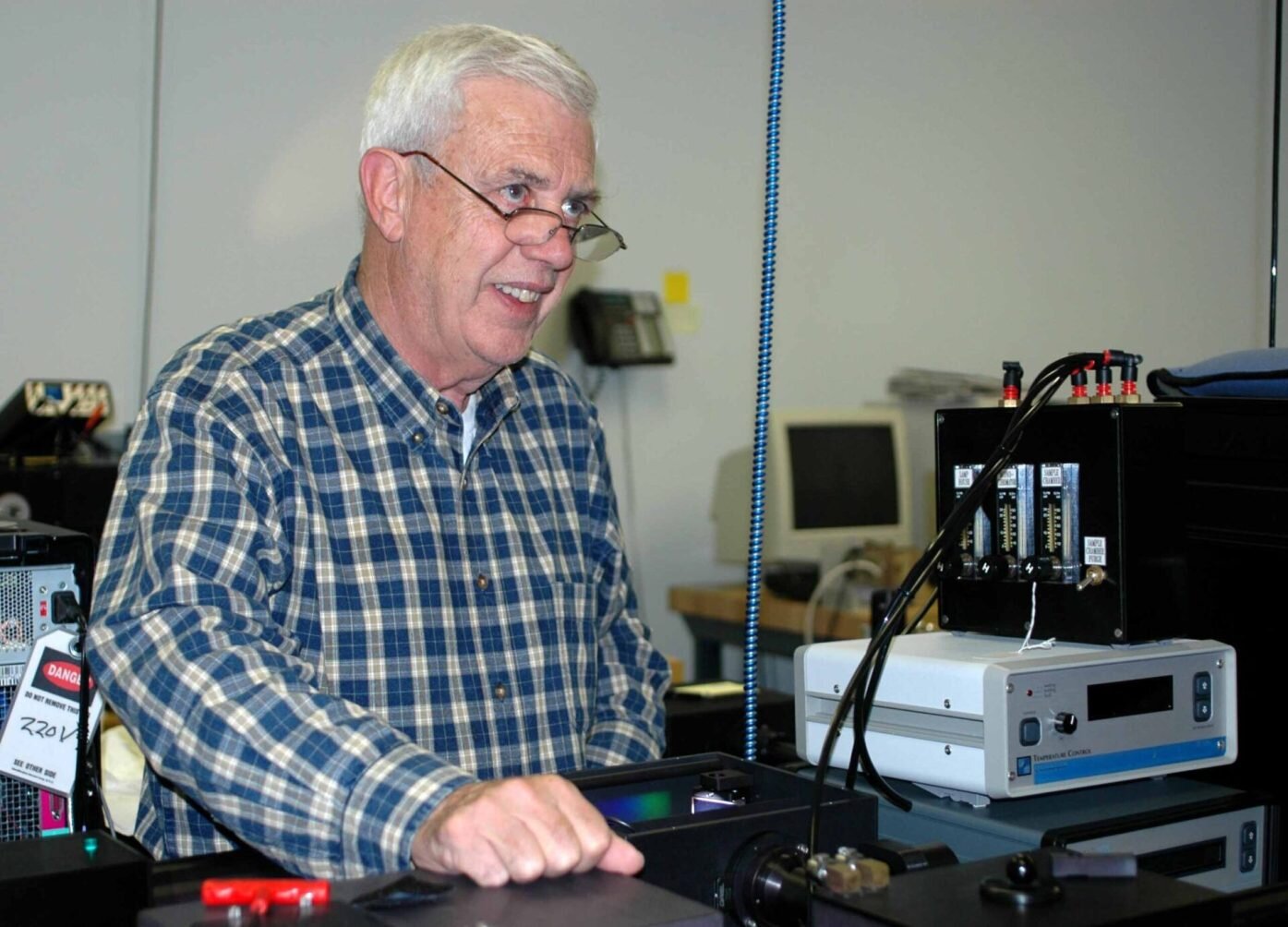
Usually, a spectrophotometer is made up of 2 instruments, specifically, a spectrometer and a photometer. A basic spectrophotometer consists of a light source, a monochromator, a collimator for straight light beam transmission, a cuvette to position a sample, and a photoelectric detector.
Not known Factual Statements About Circular Dichroism
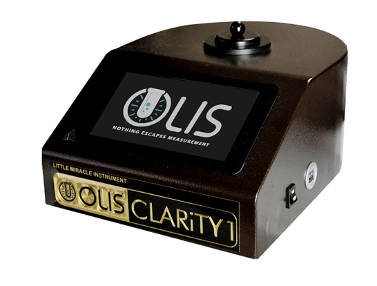
There are various kinds of spectrophotometers in different shapes and sizes, each with its own purpose or functionality. A spectrophotometer identifies just how much light is reflected by chemical elements. UV/Vis. It measures the difference in light intensity based upon the total amount of light presented to a sample and the quantity of beam that goes through the sample option
A spectrophotometer is utilized to figure out the concentration of both colorless and colored solutes in a solution. This instrument is used to identify the rate of a response.